
Freshly Printed - allow 7 days lead
Couldn't load pickup availability
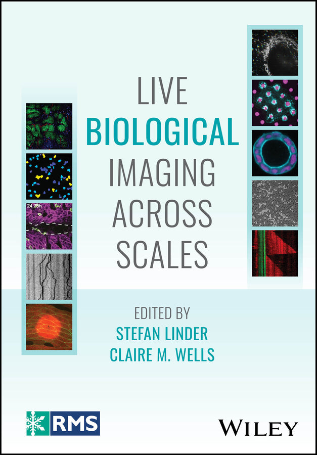
Live Biological Imaging Across Scales
Stefan Linder (Edited by), Claire M. Wells (Edited by)
9781394232383, Wiley
Hardback, published 16 April 2026
240 pages
24.9 x 17.3 x 2 cm, 0.59 kg
COMPREHENSIVE REFERENCE EXPLORING MICROSCOPY TECHNIQUES TO SUPPORT THE STUDY OF LIVING SYSTEMS WITH A UNIQUE MULTISCALE FOCUS Live Biological Imaging Across Scales presents an overview of the technologies and experimental strategies available for live imaging across scales. Each chapter highlights a different technique, working upwards from small to large scale. Readers will find critical evaluation of these techniques and perspectives on future developments in this evolving field, enabling them to effectively identify techniques that may benefit their research activities. Written by a team of leading experts and part of the Wiley-Royal Microscopical Society series, Live Biological Imaging Across Scales includes information on: Live Biological Imaging Across Scales is an essential reference for microscopists and experimental biologists, particularly those interested and/or working in the expanding field of live imaging in biology, as well as those working in the commercial microscopy industry supply and development.
List of Contributors xi Preface xv 1 Super- Resolution Imaging 1 1.1 Resolution, Widefield, and Confocal Imaging 1 1.2 SR- SIM: “Super- Resolution” Structured Illumination Using Spatially Varying Illumination 2 1.3 STED: Stimulated Emission Depletion Microscopy 5 1.4 Single Molecule Localisation Microscopies 7 1.5 Single- Molecule Light- Sheet Microscopy 11 1.6 Computation for Live- Cell Super Resolution 12 1.7 Advanced Applications in Cells 13 1.8 Concluding Remarks 14 Acknowledgments 14 References 14 2 Peering into the Septin Cytoskeleton – New Insights Unlocked by Microscopy Advances 23 2.1 Introduction – the Septin Cytoskeleton 23 2.2 Total Internal Reflection Fluorescence (TIRF) Microscopy Assays Elucidate Functions of Microtubule- and Actin- Associated Septins 25 2.3 Super- Resolution Fluorescence Microscopy Is Resolving the Nanoscale Organization of Membrane- and Actomyosin- Associated Septins 28 2.4 High- Speed Atomic Force Microscopy (HS- AFM) Reveals How Septins Assemble on Membranes 30 2.5 Cryo- Electron Tomography (Cryo- ET) Begins to Unveil the Supramolecular Organization of Septin Structures 32 2.6 Scoping the Future – Challenges and Needs of Septin Imaging 35 Acknowledgments 36 References 36 3 Imaging Phagocytosis: From Cells to Molecules 43 3.1 Introduction 43 3.2 Fluorescence Labeling to Study Phagocytosis 45 3.2.1 Antibodies to Endogenous or Epitope- Tagged Proteins 45 3.2.2 Fluorescent Chimeras of Proteins 45 3.3 Pros and Cons of Microscopy Techniques to Examine Phagocytosis 46 3.3.1 Epifluorescence and Confocal Microscopy 46 3.3.2 Lattice Light- Sheet Microscopy 46 3.3.3 Frustrated Phagocytosis 47 3.4 Single- Particle Tracking 48 3.4.1 The Principle 48 3.4.2 Requirements of SPT 49 3.4.3 Motion Types 50 3.4.4 Physicochemical and Optical Properties of Fluorescent Probes Affecting SPT 51 3.4.4.1 Specificity 51 3.4.4.2 Valency 52 3.4.4.3 Size and Molecular Weight 52 3.4.4.4 Brightness 52 3.4.4.5 Blinking 52 3.4.4.6 Photostability 52 3.4.5 Photophysical Properties of Probes Commonly Used in SPT 55 3.4.5.1 Fluorescent Proteins 55 3.4.5.2 Organic Dyes 55 3.4.5.3 Quantum Dots 56 3.4.5.4 Protein Tags 56 3.4.6 Application of Fluorescent Probes in Labeling Biomolecules for SPT 57 3.4.7 Application of SPT to Study Phagocytosis 58 3.5 Concluding Remarks 58 Acknowledgments 59 References 60 4 Imaging Wound Healing Across Scales 65 4.1 Introduction: Starting from Scratch 65 4.2 Visualizing Wound Healing in Real Time 67 4.3 Dynamics of Cell Migration During Wound Healing 69 4.3.1 Immune Cell Migration 69 4.3.2 Collective Migration of Epithelial Cells 72 4.4 ECM Remodeling 74 4.5 Conclusions and Perspectives 75 Acknowledgments 76 References 76 5 Leukocyte and Cancer Adhesion Live- Cell Imaging 81 5.1 Introduction 81 5.2 Live- Cell Imaging of Leukocyte Adhesion 82 5.3 Live- Cell Imaging of Cancer Adhesion 87 5.4 Concluding Remarks 91 References 91 6 Bridging the Gap Between In Vitro and In Vivo Studies with Live- Cell Imaging of Organoids 97 6.1 Introduction 97 6.1.1 Importance of Live Imaging to Analyze the Physiological Behavior of Cells and Tissues 97 6.1.2 Light Dose and Peak Intensity Determine Phototoxicity in the Specimen 98 6.1.3 Irradiance and Light Dose in 3D (Volumetric) Fluorescence Microscopy 100 6.1.3.1 Laser Scanning Confocal Microscopy 100 6.1.3.2 Spinning Disk Confocal Microscopy 101 6.1.3.3 Multiphoton Microscopy 101 6.1.3.4 Light- Sheet Fluorescence Microscopy 103 6.1.4 Live Imaging with WFM 105 6.1.5 Artificial Intelligence for Live Imaging 105 6.2 Organoids: A Paradigm Change Bridging the Gap Between In Vivo and In Vitro Assays 106 6.2.1 Three- Dimensional Cell Cultures 106 6.2.2 Definition of Organoid 106 6.2.3 Types of Organoids 108 6.2.4 Applications of Organoids: Bridging In Vivo and In Vitro Studies 108 6.3 Live Imaging of Organoids 109 6.3.1 The Challenging Optical Properties of Organoids 109 6.3.2 Selected Applications 110 6.3.2.1 Widefield Microscopy 110 6.3.2.2 Confocal Microscopy 111 6.3.2.3 Light- Sheet Fluorescence Microscopy 113 6.3.2.4 “Classical” LSFM 114 6.3.2.5 Single- Objective LSFM 115 6.3.2.6 Inverted (“Open- Top”) LSFM 116 6.4 Conclusion: Key Points and Future Perspectives 117 References 117 7 Tiny Organisms, Big Advantages: Exploring Membrane Damage Responses Using Dictyostelium discoideum in High- Throughput Single- Cell Analyses 123 7.1 Introduction 123 7.2 Dictyostelium discoideum, a Workhorse of Cellular Microbiology 125 7.3 Optimized Live Imaging Protocols to Track LLOMe- Mediated Endolysosomal Damage and Repair 127 7.4 Perspectives/Discussion 130 7.5 Annexes: Detailed Protocols 132 7.5.1 Annex 1: Establishment of Stable Safe- Haven Cell Lines 132 7.5.2 Annex 2: LLOMe Protocol 133 References 135 8 Live Imaging in Caenorhabditis elegans Neurons 141 8.1 Introduction 141 8.2 Worm Immobilization 141 8.3 Live Imaging in C. elegans 143 8.3.1 Imaging Actin and Cytoskeletal Elements Associated with Actin 143 8.3.2 Microtubule Imaging 147 8.3.3 Calcium Imaging 148 8.3.4 Mitochondria Imaging 149 8.3.5 Neurotransmitter Sensors 150 8.3.6 Voltage 151 8.3.7 Extracellular Vesicles 151 8.3.8 Retrograde Labeling of Endocytic Compartments 152 8.3.9 Exocytosis/Endocytosis of Vesicles 152 8.3.10 Organelle Imaging 153 8.4 Conclusion 156 Acknowledgments 156 References 156 9 It’s Just a Scratch – Lessons from Epidermal Wound Closure in Drosophila 171 9.1 From Stitches, Scratches, and Cuts 171 9.2 Drosophila as Model to Study Wound Healing 172 9.3 Calcium Bursts Orchestrate Early Wound Signaling Across Species 173 9.4 Forcing Wounds Closed – Actin Dynamics Drive Early Epidermal Wound Closure 176 9.5 Tensile Forces – Junctional Remodeling in Epidermal Wound Closure 180 9.6 Calcium Flashes Trigger the Cellular Immune Response by ROS Gradients 181 9.7 (Wound) Closing Remarks 182 Acknowledgments 182 References 183 10 Intravital Imaging of Tumors 191 10.1 Introduction 191 10.2 Capabilities of Laser Scanning Microscopy 192 10.3 Essential Components of Laser Scanning Microscopes 193 10.4 Challenges Inherent in Imaging Live Organisms 194 10.5 Techniques for Overcoming Motion 196 10.6 Unique Metrics Attainable with IVM 197 10.7 Examples of Recent Applications of IVM 199 10.7.1 Using Negative Contrast Imaging 199 10.7.2 Intravital Imaging of Lung Metastasis Using a Permanent Lung Imaging Window 200 10.7.3 Intravital Imaging of Cancer Stem Cells 202 10.8 Ongoing Challenges and New Techniques to Address Them 203 10.8.1 Limits to Penetration Depth 203 10.8.2 Limits to the Number of Simultaneous Labels 203 10.9 Conclusion 205 References 205 Index 211
Alistair Curd, Amir Rahmani, Oliver Umney, Aleks Ponjavic, and Michelle Peckham
Andrew W. Schaefer and Elias T. Spiliotis
Trieu Le, Ava Kavianpour, Spencer Freeman, and Sergio Grinstein
Simran Rawal and Tamal Das
Camilla Cerutti
Francesco Pampaloni
Angélique Perret, Céline Michard, Dimitri Moreau, and Thierry Soldati
Badal Singh Chauhan and Sandhya P. Koushika
Dennis Klug and Sven Bogdan
Yookyung Jung and David Entenberg
Subject Areas: Chemistry [PN]