
Freshly Printed - allow 7 days lead
Couldn't load pickup availability
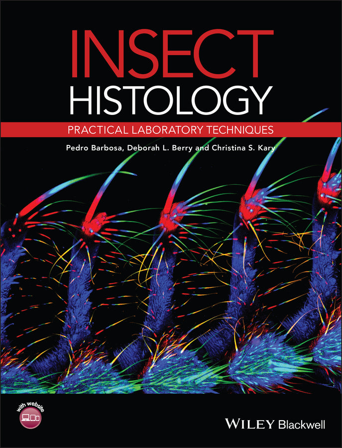
Insect Histology
Practical Laboratory Techniques
Pedro Barbosa (Author), Deborah Berry (Author), Christina K. Kary (Author)
9781444336962, Wiley
Paperback / softback, published 21 November 2014
368 pages
24.4 x 18.5 x 2.3 cm, 0.862 kg
“A very useful book to have in your library.” (British Journal of Entomology & Natural History, 1 March 2015)
This helpful guide is self-contained, holding all of the information you will need for the preparation of histological slides of all types of insect tissues. It explains the essential procedures for their preparation, providing step-by-step information on each phase, for example, on fixation, dehydrating, clearing, embedding, and staining. The laboratory manual explains the cutting-edge methodology carried out today, and provides a thorough collection of both historically established and modern insect histology techniques side-by-side. It is the first book to present these various techniques appropriate to insects. Key issues covered include: Photographs show the likely results of procedures, components of the process, and advantages of the use of particular approaches or compounds. As a comprehensive resource, this book is for students and researchers in entomology, systematics, developmental biology, insect cell biology, and morphology, wanting a clear introduction to the procedures for the histological display of insect tissues.
Preface ix Acknowledgements xi Introduction xiii About the companion website xiv 1 Problems of sclerotized chitin: Softening insect cuticle 1 1.1 Introduction 1 1.2 General Methods 3 1.3 Preparations of insect eggs 14 1.4 Double Embedding Techniques 16 References 19 2 Fixation 21 2.1 Introduction 21 2.2 Aldehyde based fixatives 21 2.3 Protein denaturing 30 2.4 Picric acid based 33 2.5 Mercuric chloride based 37 2.6 SEM/TEM 40 2.7 Other 46 References 51 3 Dehydrating, clearing, and embedding 54 3.1 Dehydration 54 3.2 Clearing 60 3.3 Embedding General 65 3.4 Embedding – Ester Wax 73 3.5 Embedding – Methacrylate 74 References 77 4 Staining 79 4.1 Single-contrast staining – Carmines 81 4.2 Single contrast staining – Nuclear Stains 83 4.3 Single contrast staining – General Stains 86 4.4 Single contrast staining – Golgi 89 4.5 Single contrast staining – Eggs 89 4.6 Single contrast staining – Silver Stains 90 4.7 Polychrome staining techniques – General 92 4.8 Polychrome staining – Brain/Nerve 102 4.9 Polychrome staining – blood 103 4.10 Single contrast procedures for chitinous material 105 4.11 Polychrome staining procedures for chitinous material 106 4.12 Polychrome staining for chitinous material – KOH 110 4.13 Polychrome staining for chitinous material – Differential staining of Individual Organs 111 4.14 Staining of specific tissues 113 4.15 Two dye combinations 114 References 117 5 Immunohistochemical techniques 119 5.1 Introduction 119 5.2 General immunostaining techniques 127 5.3 Immunolabeling of samples for Transmission Electron Microscopy (TEM) 135 5.4 Proliferation assays 140 5.5 Methods to detect specific proteins 142 References 144 6 Use of genetic markers in insect histology 146 6.1 Introduction 146 6.2 Inducible genetic markers 149 6.3 Mosaic gene expression 156 6.4 Fluorescent markers for live imaging and kinetic microscopy 165 References 169 7 Fluorescence 171 7.1 Introduction 171 References 192 8 Mounting 194 8.1 Introduction 194 References 206 9 Preparation of whole mounts 208 9.1 Introduction 208 References 229 10 Preparation of whole mounts for staining 231 10.1 Introduction 231 10.2 Detection of NAPDHd 237 10.3 SEM 238 10.4 In situ hybridization 240 References 244 11 Preparation of genitalia, mouthparts and other body parts 246 References 256 12 Preparation of chromosomes 258 References 288 13 Preparation of other specific insect organs and tissues 290 13.1 Introduction 290 References 323 Appendix Dissecting fluids and saline solutions 325 Index 333
Subject Areas: Veterinary medicine [MZ]