
Freshly Printed - allow 7 days lead
Couldn't load pickup availability
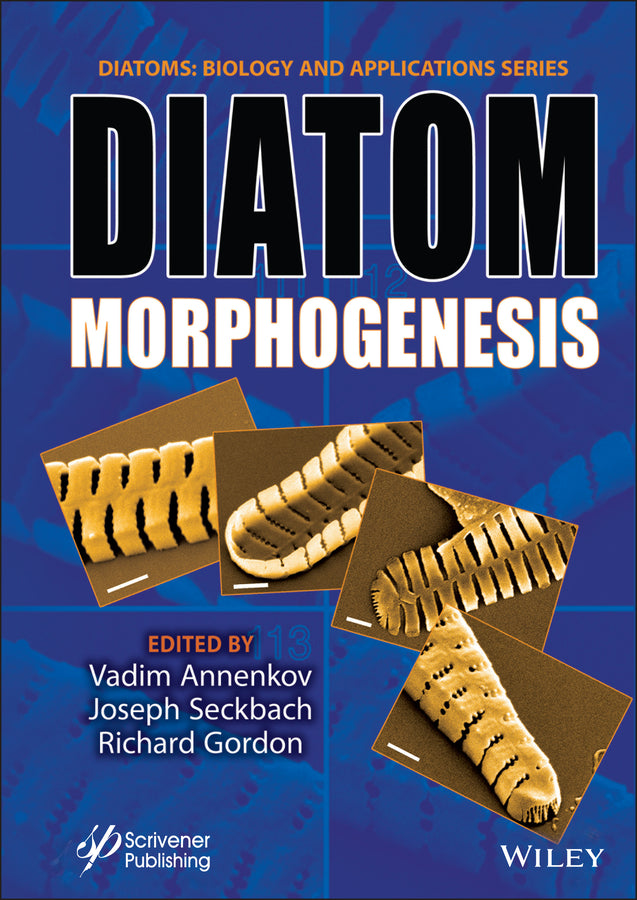
Diatom Morphogenesis
Vadim V. Annenkov (Edited by), Joseph Seckbach (Edited by), Richard Gordon (Edited by)
9781119487951, Wiley
Hardback, published 17 December 2021
448 pages
1 x 1 x 1 cm, 0.454 kg
DIATOM MORPHOGENESIS A unique book presenting the range of silica structures formed by diatoms, theories and hypotheses of how they are made, and applications to nanotechnology by use or imitation of diatom morphogenesis. There are up to 200,000 species of diatoms, each species of these algal cells bearing an ornate, amorphous silica glass shell. The silica is structured at 7 orders of magnitude size range and is thus the most complex multiscalar solid structure known. Recent research is beginning to unravel how a single cell marshals chemical, physical, biochemical, genetic, and cytoskeletal processes to produce these single-cell marvels. The field of diatom nanotechnology is advancing as this understanding matures. Diatoms have been actively studied over the recent 10-20 years with various modern equipment, experimental and computer simulation approaches, including molecular biology, fluorescence-based methods, electron, confocal, and AFM microscopy. This has resulted in a huge amount of information but the key stages of their silica morphogenesis are still not clear. This is the time to reconsider and consolidate the work performed so far and to understand how we can go ahead. The main objective of this book is to describe the actual situation in the science of diatom morphogenesis, to specify the most important unresolved questions, and to present the corresponding hypotheses. The following areas are discussed: Audience Researchers, scientists, and graduate students in the fields of phycology, general biology, marine sciences, the chemistry of silica, materials science, and ecology.
Preface xv Part 1: General Issues 1 1 Introduction for a Tutorial on Diatom Morphology 3 1.1 Diatoms in Brief 3 1.2 Tools to Explore Diatom Frustule Morphology 7 1.3 Diatom Frustule 3D Reconstruction 12 1.3.1 Recommended Steps to Understand the Complex Diatom Morphology: A Guide for Beginners 13 1.4 Conclusion 15 Acknowledgements 15 References 15 2 The Uncanny Symmetry of Some Diatoms and Not of Others: A Multi-Scale Morphological Characteristic and a Puzzle for Morphogenesis 19 2.1 Introduction 20 2.1.1 Recognition and Symmetry 21 2.1.2 Symmetry and Growth 24 2.1.3 Diatom Pattern Formation, Growth, and Symmetry 25 2.1.4 Diatoms and Uncanny Symmetry 27 2.1.5 Purpose of This Study 28 2.2 Methods 28 2.2.1 Centric Diatom Images Used for Analysis 28 2.2.2 Centric Diatoms, Morphology, and Valve Formation 34 2.2.3 Image Entropy and Symmetry Measurement 36 2.2.4 Image Preparation for Measurement 37 2.2.5 Image Tilt and Slant Measurement Correction for Entropy Values 38 2.2.6 Symmetry Analysis 39 2.2.7 Entropy, Symmetry, and Stability 40 2.2.8 Randomness and Instability 42 2.3 Results 43 2.3.1 Symmetry Analysis 43 2.3.2 Valve Formation—Stability and Instability Analyses 49 2.4 Discussion 51 2.4.1 Symmetry and Scale in Diatoms 55 2.4.2 Valve Formation and Stability 56 2.4.3 Symmetry, Stability and Diatom Morphogenesis 57 2.4.4 Future Research—Symmetry, Stability and Directionality in Diatom Morphogenesis 58 References 59 3 On the Size Sequence of Diatoms in Clonal Chains 69 3.1 Introduction 70 3.2 Mathematical Analysis of t he Size Sequence 73 3.2.1 Alternative Method for Calculating the Size Sequence 73 3.2.2 Self-Similarity and Fractal Structure 75 3.2.3 Matching Fragments to a Generation Based on Known Size Indices of the Fragment 76 3.2.4 Sequence of the Differences of the Size Indices 78 3.2.5 Matching Fragments to a Generation Based on Unknown Size Indices of the Fragment 80 3.2.6 Synchronicity of Cell Divisions 81 3.3 Observations 82 3.3.1 Challenges in Verifying the Sequence of Sizes 82 3.3.2 Materials and Methods 83 3.3.3 Investigation of the Size Sequence of a Eunotia sp. 84 3.3.4 Synchronicity 86 3.4 Conclusions 87 Acknowledgements 88 Appendix 3A L-System for the Generation of the Sequence of Differences in Size Indices of Adjacent Diatoms 88 Appendix 3B Probability Consideration for Loss of Synchronicity 89 References 91 4 Valve Morphogenesis in Amphitetras antediluviana Ehrenburg 93 4.1 Introduction 93 4.2 Material and Methods 94 4.3 Observations 94 4.3.1 Amphitetras antediluviana Mature Valves 94 4.3.2 Amphitetras antediluviana Forming Valves 96 4.3.3 Amphitetras antediluviana Girdle Band Formation 101 4.4 Conclusion 101 Acknowledgments 102 References 102 Glossary 104 Part 2: Simulation 105 5 Geometric Models of Concentric and Spiral Areola Patterns of Centric Diatoms 107 5.1 Introduction 107 5.2 Set of Common Rules Used in the Models 109 5.3 Concentric Pattern of Areolae 109 5.4 Spiral Patterns of Areolae 110 5.4.1 Unidirectional Spiral Pattern 111 5.4.2 Bidirectional Spiral Pattern 113 5.4.3 Common Genesis of Unidirectional and Bidirectional Spiral Patterns 113 5.5 Conversion of an Areolae-Based Model Into a Frame-Based Model 114 5.6 Conclusion 114 Acknowledgements 114 References 115 6 Diatom Pore Arrays’ Periodicities and Symmetries in the Euclidean Plane: Nature Between Perfection and Imperfection 117 6.1 Introduction 118 6.2 Materials and Methods 122 6.2.1 Micrograph Segmentation 123 6.2.2 Two-Dimensional Fast Fourier Analysis and Autocorrelation Function Analysis 123 6.2.3 Lattice Measurements and Recognition 123 6.2.4 Accuracy of 2D ACF-Based Calculations 125 6.2.5 The Perfection of the Unit Cell Parameters Between Different Parts (Groups of Pore Arrays) of the Same Valve and the Same Micrograph 126 6.3 Results and Discussion 126 6.3.1 Toward Standardization of the Methodology for the Recognition of 2D Periodicities of Pore Arrays in Diatom Micrographs 126 6.3.1.1 Using Two-Dimensional Fast Fourier Transform Analysis 126 6.3.1.2 Using Two-Dimensional Autocorrelation Function 131 6.3.1.3 The Accuracy of Lattice Parameters’ Measurements Using the Proposed 2D ACF Analysis 134 6.3.2 Exploring the Periodicity in Our Studied Micrographs and the Possible Presence of Different Types of 2D Lattices in Diatoms 137 6.3.2.1 Irregular Pore Scattering (Non-Periodic Pores) 137 6.3.2.2 Linear Periodicity of Pores in Striae (1D Periodicity) 138 6.3.2.3 The Different 2D Lattices in Diatom Pore Arrays 140 6.3.3 How Perfectly Can Diatoms Build Their 2D Pore Arrays? 146 6.3.3.1 Variation of the 2D Lattice Within the Connected Pore Array of the Valve 146 6.3.3.2 Comparison of 2D Lattice Parameters and Degree of Perfection of Distinct Pore Array Groups in the Same Micrograph and Valve but With Different Rotational or Reflection Symmetry 148 6.3.3.3 The Perfection of 2D Lattices of Diatom Pore Arrays Compared to Perfect (Non-Oblique) 2D Bravais Lattices 148 6.3.4 Planar Symmetry Groups to Describe the Whole Diatom Valve Symmetries and Additionally Describe the Complicated 2D Periodic Pore Arrays’ Symmetries 149 6.3.4.1 Rosette Groups 150 6.3.4.2 Frieze Groups 151 6.3.4.3 Wallpaper Groups 153 6.4 Conclusion 153 Acknowledgment 154 Glossary 154 References 155 7 Quantified Ensemble 3D Surface Features Modeled as a Window on Centric Diatom Valve Morphogenesis 159 7.1 Introduction 159 7.1.1 From 3D Surface Morphology to Morphogenesis 160 7.1.2 Geometric Basis of 3D Surface Models and Analysis 163 7.1.3 Differential Geometry of 3D Surface 163 7.1.4 3D Surface Feature Geometry and Morphological Attributes 165 7.1.5 Centric Diatom Taxa Used as Exemplars in 3D Surface Models for Morphogenetic Analysis 166 7.1.6 Morphogenetic Descriptors of Centric Diatoms in Valve Formation as Sequential Change in 3D Surface Morphology 166 7.1.7 Purposes of This Study 167 7.2 Methods 168 7.2.1 Measurement of Ensemble Surface Features and 3D Surface Morphology: Derivation and Solution of the Jacobian, Hessian, Laplacian, and Christoffel Symbols 168 7.2.1.1 The Jacobian of 3D Surface Morphology 168 7.2.1.2 Monge Patch 169 7.2.1.3 First and Second Fundamental Forms and Surface Characterization of the Monge Patch 169 7.2.1.4 3D Surface Characterization via Gauss and Weingarten Maps and the Fundamental Forms 170 7.2.1.5 Peaks, Valleys, and Saddles of Surface Morphology and the Hessian 170 7.2.1.6 Smoothness as a Characterization of Surface Morphology and the Laplacian 171 7.2.1.7 Point Connections of 3D Surface Morphology and Christoffel Symbols 171 7.2.1.8 Protocol for Using Centric Diatom 3D Surface Models and Their Ensemble Surface Features in Valve Formation Analysis 173 7.3 Results 174 7.4 Discussion 184 7.4.1 Ensemble Surface Features and Physical Characteristics of Valve Morphogenesis 186 7.4.2 Factors Affecting Valve Formation 187 7.4.3 Diatom Growth Patterns—Buckling and Wave Fronts 187 7.4.4 Valve Formation, Ensemble Surface Features, and Self-Similarity 189 7.4.5 Diatom Morphogenesis: Cytoplasmic Inheritance and Phenotypic Plasticity 189 7.4.6 Phenotypic Variation and Ensemble Surface Features: Epistasis and Canalization 190 7.5 Conclusions 190 Acknowledgment 191 References 191 8 Buckling: A Geometric and Biophysical Multiscale Feature of Centric Diatom Valve Morphogenesis 195 8.1 Introduction 196 8.2 Purpose of Study 197 8.3 Background: Multiscale Diatom Morphogenesis 198 8.3.1 Valve Morphogenesis—Schemata of Schmid and Volcani and of Hildebrand, Lerch, and Shrestha 198 8.3.2 Valve Formation—An Overview at the Microscale 199 8.3.3 Valve Formation—An Overview at the Meso- and Microscale 200 8.3.4 Valve Formation—An Overview at the Meso- and Nanoscale 200 8.4 Biophysics of Diatom Valve Formation and Buckling 201 8.4.1 Buckling as a Multiscale Measure of Valve Formation 201 8.4.2 Valve Formation—Cytoplasmic Features and Buckling 202 8.4.3 Buckling: Microtubule Filaments and Bundles 203 8.4.4 Buckling: Actin Filament Ring 204 8.5 Geometrical and Biophysical Aspects of Buckling and Valve Formation 205 8.5.1 Buckling: Geometry of Valve Formation as a Multiscale Wave Front 205 8.5.2 Buckling: Valve Formation and Hamiltonian Biophysics 207 8.5.3 Buckling: Valve Formation and Deformation Gradients 208 8.5.4 Buckling: Multiscale Measurement With Respect to Valve Formation 210 8.5.5 Buckling: Krylov Methods and Association of Valve Surface Buckling With Microtubule and Actin Buckling 210 8.6 Methods 211 8.6.1 Constructing and Analyzing 3D Valve Surface and 2D Microtubule and Actin Filament Models 211 8.6.2 Krylov Methods: Associating Valve Surface With Microtubule and Actin Filament Buckling 212 8.7 Results 212 8.8 Conclusion 216 References 223 9 Are Mantle Profiles of Circular Centric Diatoms a Measure of Buckling Forces During Valve Morphogenesis? 231 9.1 Introduction 231 9.2 Methods 233 9.2.1 Background: Circular Centric 2D Profiles and 3D Surfaces of Revolution 236 9.3 Results 238 9.3.1 Approximate Constant Profile Length Representing Approximate Same Sized Valves 239 9.3.2 Change in Profile Length Representing Size Reduction During Valve Morphogenesis 240 9.3.2.1 Inferences About Complementarity and Heterovalvy 242 9.3.3 Are Profiles Measures of Buckling Forces During Valve Morphogenesis? 243 9.4 Discussion 245 9.4.1 Laminated Structures and Mantle Buckling Forces Affecting the Valve Profile 247 9.5 Conclusion 248 Acknowledgement 248 References 248 Part 3: Physiology, Biochemistry and Applications 251 10 The Effect of the Silica Cell Wall on Diatom Transport and Metabolism 253 Publications by and about Mark Hildebrand 254 11 Diatom Plasticity: Trends, Issues, and Applications on Modern and Classical Taxonomy, Eco‑Evolutionary Dynamics, and Climate Change 261 11.1 Introduction 261 11.2 Model Species: Phaeodactylum tricornutum 262 11.3 Transformation Mechanisms of P. tricornutum 263 11.4 Future Advances in the Phenotypic Plasticity on P. tricornutum 263 11.4.1 Genomic and Molecular Mechanisms in Diatom Phenotypic Plasticity 263 11.4.2 Biogeography of Diatoms 263 11.4.3 Eco-Evolutionary Dynamics Approach on Diatoms Phenotypic Plasticity 264 11.4.4 Adaptive Behavior and Evolutionary Changes in Diatoms Linking to Diatom Plasticity 265 11.4.5 Climate Change and Phenotypic Plasticity 265 11.5 Conclusion 265 References 265 12 Frustule Photonics and Light Harvesting Strategies in Diatoms 269 12.1 Introduction 270 12.2 Light Spectral Characteristics and Signaling 274 12.2.1 Variation of Light Regimes 274 12.2.2 Light Perception and Signaling 275 12.3 Photosynthesis and Photo-Protection in Diatoms 276 12.3.1 Pigment-Based Light Absorption 276 12.3.2 Molecular Photo-Protection Mechanisms 276 12.3.3 Intracellular Structural Adaptation in Response to Light 277 12.3.4 Motility as a Unique Photo-Protection Mechanism 278 12.4 Frustule Photonics Related to Diatom Photobiology 279 12.4.1 An Extracellular Structure With Optical Properties 279 12.4.2 Intraspecific and Intra-Individual Variation of Frustule Periodicity 281 12.4.3 Photonic Crystal Properties 281 12.4.4 Light Confinement and Focusing 282 12.4.5 Scattering and Dispersion of Light 283 12.4.6 Attenuation of UV Light for Photo-Protection 283 12.5 Frustule Photonics in Light of Niche Differentiation 285 12.6 Conclusion 291 References 292 13 Steps of Silicic Acid Transformation to Siliceous Frustules: Main Hypotheses and Discoveries 301 13.1 Introduction 301 13.2 Penetration of the Boundary Layer: The Diatom as an Antenna for Silica 303 13.3 Getting Past the Cloud of Extracellular Material 304 13.4 Adsorption of Silica Onto the Outer Organic Coat of the Diatom 305 13.5 Getting Past the Silica Frustule or Through Its Pores 306 13.6 Getting Past the Inner Organic Coat, the Diatotepum 306 13.7 Transport of Silica Across the Cell Membrane 307 13.8 Cytoplasm Storage and Trafficking of Silica to the Places of Synthesis of the Frustule Parts 309 13.9 Transport and Patterning of Silica Across the Silicalemma 311 13.10 Precipitation and Morphogenesis of the Nascent Valve Within the Silicalemma 314 13.11 Thickening of the Valve Within the Silicalemma 319 13.12 Exteriorization of the Valve 321 13.13 Future Work Needed 321 13.14 Conclusion 323 References 326 14 The Effects of Cytoskeletal Inhibitors on Diatom Valve Morphogenesis 349 14.1 Introduction 349 14.2 Cytoskeleton and Its Role in Cell Morphogenesis 350 14.3 Abnormalities of Diatom Valve Morphogenesis Induced by Cytoskeleton Inhibitors 352 14.4 Conclusion 358 Acknowledgment 360 References 360 15 Modeling Silicon Pools in Diatoms Using the Chemistry Toolbox 365 15.1 Diatoms 365 15.2 “Silicon Pools” Biology 366 15.3 Silica Particle Formation From Silicic Acid 366 15.4 Stabilization of “Soluble” Silica Species (Monosilicic and Disilicic Acids) 370 15.4.1 Cationic Polymers 370 15.4.2 Neutral (Uncharged) Polymers 372 15.4.3 Zwitterionic Polymers 373 15.4.4 Blends of Cationic/Anionic Polymers 375 15.5 Chemical Mechanisms 376 15.6 Conclusions/Perspectives 377 Acknowledgments 378 References 378 16 The Mesopores of Raphid Pennate Diatoms: Toward Natural Controllable Anisotropic Mesoporous Silica Microparticles 383 16.1 Introduction 384 16.2 Morphology and Very Fine Ultrastructure of Diatom Frustules 386 16.3 Synthetic Mesoporous Silica 391 16.4 The Potential of Raphid Pennates’ Mesoporous Bio-Silica, Similarities, and Dissimilarities Compared With Synthetic MSM/Ns 393 16.4.1 The Current Potential of Diatom Porous Silica in Applications 393 16.4.2 Why Should We Be Interested in the Mesoporous Silica of Raphid Pennate Frustules if the Frustules of Other Species With Larger Pores Work? 393 16.4.3 Similarities and Dissimilarities Compared With Synthetic MSM/Ns 394 16.5 Our Ability to Control the Diatom Frustule’s Ultrastructure 396 16.5.1 Physicochemical Parameters Alteration Approach 397 16.5.2 Genetic Engineering Approach 398 16.6 Conclusion 399 Acknowledgment 399 References 399 Glossary 408 Index 411
Kalina Manoylov and Mohamed Ghobara
Janice L. Pappas, Mary Ann Tiffany and Richard Gordon
Thomas Harbich
Mary A. Tiffany and Bonnie L. Hurwitz
Anton M. Lyakh
Mohamed M. Ghobara, Mary Ann Tiffany, Richard Gordon and Louisa Reissig
Janice L. Pappas
Janice L. Pappas and Richard Gordon
Janice L. Pappas and Richard Gordon
Mark Hildebrand
Lawrence Victor D. Vitug
Johannes W. Goessling, Yanyan Su, Michael Kühl and Marianne Ellegaard
Vadim V. Annenkov, Elena N. Danilovtseva and Richard Gordon
Yekaterina D. Bedoshvili and Yelena V. Likhoshway
Argyro Spinthaki and Konstantinos D. Demadis
Mohamed M. Ghobara, Richard Gordon and Louisa Reissig
Subject Areas: Science: general issues [PD]