
Freshly Printed - allow 7 days lead
Couldn't load pickup availability
Diatom Microscopy
Nirmal Mazumder (Edited by), Richard Gordon (Edited by)
9781119711537, Wiley
Hardback, published 28 August 2022
368 pages
1 x 1 x 1 cm, 0.454 kg
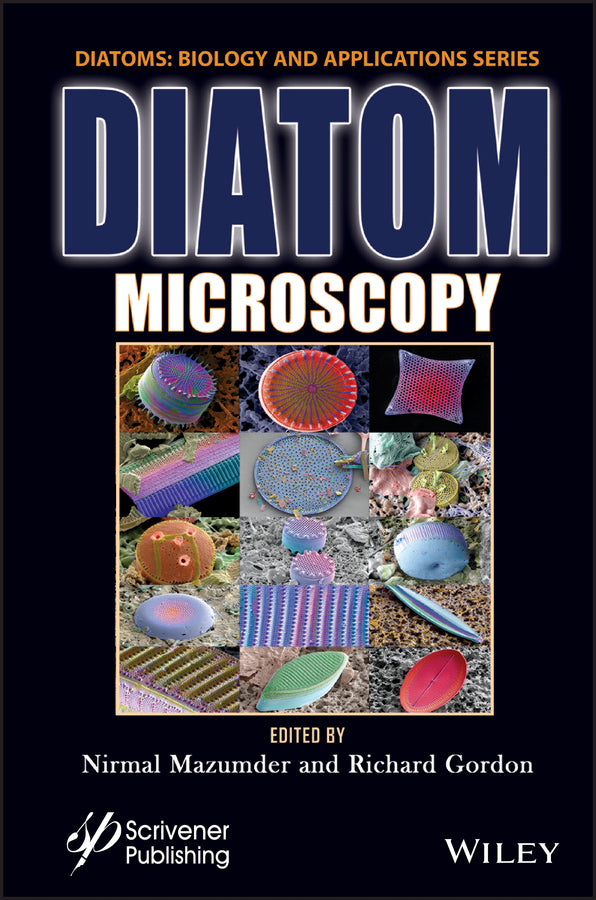
DIATOM MICROSCOPY The main goal of the book is to demonstrate the wide variety of microscopy methods being used to investigate natural and altered diatom structures. This book on Diatom Microscopy gives an introduction to the wide panoply of microscopy methods being used to investigate diatom structure and biology, marking considerable advances in recent technology including optical, fluorescence, confocal and electron microscopy, surface-enhanced Raman spectroscopy (SERS), atomic force microscopy (AFM) and spectroscopy as applied to diatoms. Each chapter includes a tutorial on a microscopy technique and reviews its applications in diatom nanotechnology and diatom research. The number of diatomists, diatom research, and their publications are increasing rapidly. Although many books have dealt with various aspects of diatom biotechnology, nanotechnology, and morphology, to our knowledge, no volume exists that summarizes advanced microscopic approaches to diatoms. Audience The intended audience is academic and industry researchers as well as graduate students working on diatoms and diatom nanotechnology, including biosensors, biomedical engineering, solar panels, batteries, drug delivery, insect control, and biofuels.
Preface xi 1 Investigation of Diatoms with Optical Microscopy 1 1.1 Introduction 2 1.2 Light Microscopy 4 1.2.1 Phase Contrast Microscopy 4 1.2.2 Differential Interference Contrast (DIC) Microscopy 6 1.2.3 Darkfield Microscopy 9 1.3 Fluorescence Microscopy 10 1.4 Confocal Laser Scanning Microscopy 12 1.5 Multiphoton Microscopy 19 1.6 Super-Resolution Optical Microscopy 22 1.7 Conclusion 25 Acknowledgement 25 References 25 2 Nanobioscience Studies of Living Diatoms Using Unique Optical Microscopy Systems 33 Abbreviations 33 2.1 Trajectory Analysis of Gliding Among Individual Diatom Cells Using Microchamber Systems 35 2.2 Direct Observation of Floating Phenomena of Individual Diatoms Using a “Tumbled” Microscope System 40 2.3 Three-Dimensional Physical Imaging of Living Diatom Cells Using a Holographic Microscope System 44 Acknowledgements 47 References 47 3 Recent Insights Into the Ultrastructure of Diatoms Using Scanning and Transmission Electron-Microscopy 57 3.1 Introduction 58 3.2 Scanning Electron Microscopy (SEM) of Diatoms 58 3.3 Transmission Electron Microscopy (TEM) of Diatoms 65 3.3.1 Limitations 69 3.4 Conclusion 73 References 73 4 Atomic Force Microscopy Study of Diatoms 81 4.1 Introduction 82 4.2 Types of AFM Modes 86 4.3 Sample Preparation and Methods 88 4.4 Study of Diatom Ultrastructure Under AFM 88 4.5 Conclusion 102 Glossary 104 Acknowledgement 104 References 105 5 Refractive Index Tomography for Diatom Analysis 111 5.1 Introduction 112 5.2 Fundamentals of PC-ODT 113 5.3 Experimental Setup for PC-ODT 117 5.4 Diatom RI Reconstructions with Bright-Field Illumination 120 5.5 Illumination Impact on PC-ODT Performance 126 5.6 Concluding Remarks 131 Acknowledgement 134 References 134 6 Luminescent Diatom Frustules: A Review on the Key Research Applications 139 6.1 Introduction 140 6.2 Key Research Applications of Luminescence Properties of Diatom Frustules 141 6.2.1 Novel Nanophotonic and Optoelectronic Applications of Luminescent Diatom Frustules 142 6.2.2 Applications of Diatom Luminescence in Sensing 145 6.2.3 Biomedical Applications of Diatom Luminescence 149 6.2.4 Other Studies on Diatom Luminescence 151 6.3 Future Perspectives 167 6.4 Conclusion 168 Acknowledgement 168 References 168 7 Micro to Nano Ornateness of Diatoms from Geographically Distant Origins of the Globe 179 7.1 Introduction 180 7.2 Materials and Methods 183 7.2.1 Diatom Samples and Microscopy 183 7.2.1.1 By Michael J. Stringer 183 7.2.1.2 Diatom Oamaru Slides by Diane Winter 184 7.2.1.3 By Daniel Mathys 184 7.2.1.4 Diatom Sampling, Slide Preparation and Imaging from Himalayas, Plains and Arabian Sea, India 185 7.3 Diatoms from Different Geographical Origins of the World 185 7.3.1 Oamaru Diatoms 185 7.3.2 Diatom Images Gifted by Michael J. Stringer 186 7.3.3 Diatoms from Natural History Museum Basel, Switzerland a Piece of Art by Daniel Mathys 190 7.3.4 Diatoms from India 193 7.4 Conclusion 216 7.5 Acknowledgements 216 References 216 8 Types of X-Ray Techniques for Diatom Research 221 8.1 Introduction 221 8.2 Applications 222 8.2.1 Synchrotron Radiation-Based X-Ray Techniques 222 8.2.2 X-Ray Computed Tomography 224 8.2.3 X-Ray Fluorescence-Based Techniques 226 8.2.4 X-Ray Microanalysis 227 8.2.5 X-Ray Absorption-Based Techniques 228 8.2.6 X-Ray Diffraction 229 8.2.7 Other X-Ray-Based Techniques 230 8.3 Conclusions 233 Glossary 233 References 234 9 Diatom Assisted SERS 237 9.1 Introduction 237 9.2 Diatom 239 9.2.1 Basic Overview 239 9.2.2 Physiological Characteristics 239 9.2.3 Optical and Relevant Properties 240 9.3 Raman Scattering 241 9.3.1 Basics 241 9.3.2 Surface Enhanced Raman Scattering 242 9.3.3 Optoelectronic Investigations 242 9.4 SERS Through Diatom: Fundamentals and Application Overview 243 9.5 Conclusion and Future Outlook 245 References 246 10 Diatoms as Sensors and Their Applications 251 10.1 Introduction 251 10.2 Diatoms as Biosensors 255 10.2.1 Electrochemical Sensors 260 10.2.2 Plasmonic Sensors 261 10.2.3 Immunoassay Sensors 264 10.2.4 Optical and Optofluidic Sensors 268 10.2.5 Biochemical Sensors 271 10.2.6 FRET-Based Sensors 273 10.2.7 Microfluidics-Based Sensors 274 10.3 Conclusion 276 Acknowledgments 276 References 277 11 Diatom Frustules: A Transducer Platform for Optical Detection of Molecules 283 11.1 Introduction 284 11.2 Optical Properties of Diatom Frustules 285 11.2.1 Diatom as a Photoluminescent Materials 285 11.2.2 Diatom as a Photonic Crystal 287 11.2.3 Diatoms as a SERS Substrate 288 11.3 Methods Involved in Thin Film Deposition of Diatom Frustules 290 11.4 Diatom as an Optical Transducer for Biosensors 294 11.5 Diatom as an Optical Transducer for Gas/Chemical Sensors 297 11.6 Conclusion 300 References 301 12 Effects of Light on Physico-Chemical Properties of Diatoms 307 12.1 Introduction 308 12.2 Effect of Light on Diatom Function and Morphology 310 12.2.1 Effect of Light Intensity on Diatom Morphology 310 12.2.2 Effect of Light Intensity on Diatom Growth 313 12.2.3 Effect of Light Intensity on Photosynthesis in Diatoms 318 12.2.4 Effect of Wavelength of Light on Diatom Pigment System 321 12.2.5 Effect of Light Intensity on the Physiology of Diatoms 327 12.3 Conclusion 330 Acknowledgment 330 References 330 Index 335
Shih-Ting Lin, Ming-Xin Lee and Guan-Yu Zhuo
Kazuo Umemura
Dharshini Gopal, Shweta Chakrabarti, Dataram Venkata Srikar Keshav, Richard Gordon and Nirmal Mazumder
Ishita Chakraborty, Shweta Chakrabarti, Vishwanath Managuli and Nirmal Mazumder
Juan M. Soto, José A. Rodrigo and Tatiana Alieva
Jayur Tisso, Shruthi Shetty, Nirmal Mazumder, Ankur Gogoi and Gazi A. Ahmed
Mohd Jahir Khan, Daniel Mathys and Vandana Vinayak
Mridula Sunder, Neha Acharya, Smitha Nayak, Richard Gordon and Nirmal Mazumder
Rajib Biswas and Sankar Biswas
Priyasha De and Nirmal Mazumder
Viji S., Ponpandian N. and Viswanathan C.
Janardan Sen, Priyal Dhawan, Priyasha De and Nirmal Mazumder
Subject Areas: Biology, life sciences [PS]