
Freshly Printed - allow 7 days lead
Couldn't load pickup availability
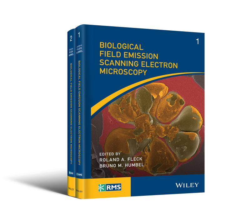
Biological Field Emission Scanning Electron Microscopy, 2 Volume Set
Roland A. Fleck (Edited by), Bruno M. Humbel (Edited by)
9781118654064, Wiley
Hardback, published 26 April 2019
752 pages
26.9 x 18.8 x 5.1 cm, 2.2 kg
Since the first commercial SEM (scanning electron microscope) was produced by Cambridge Instruments in 1965, the resolution,image contrast, and operability have improved dramatically which have turned field emission SEM into an increasingly valuable tool in the life science community. The challenge of life science microscopy is to provide the structural information for the correlation of structure and function in complex biological systems. For this reason, this two-volume edition comes as a welcome addition to the biologist’s library, since they encompass all the information needed to approach life science FEG SEM imaging challenges, including detailed descriptions of the instrumentation and analytical techniques currently available in the field. There are many features of this edition that make it a useful resource for both the beginner and more advanced microscopist. First, the two volumes are composed of 31 self-contained chapters, each written by leading authorities on these subjects. Secondly, the chapters are richly illustrated and most chapters are complemented with a comprehensive, valuable, and up-to-date list of references. Accordingly, the user who seeks to purchase new equipment or select the appropriate technique will find useful information in both volumes. In the initial nine chapters of vol. 1, the reader is given a comprehensive historical review and introduction to the possibilities of the current technology—choice of microscopy method and specimen preparation as well as cryo and room temperature options—edited by representatives from the major companies in the field. The three last chapters of vol. 1 are dedicated to a review of specimen preparation methods—chemical fixation and cryo methods—which are a must-read for any scientist in this field. In vol. 2 in the first five chapters, the reader is introduced to more special applications and results of the techniques, spanning from pathology and infectious diseases, plant cell wall and nuclear envelope, yeast cell to food research. The three following chapters focus on cryo-FEGSEM in biology, preparation protocols of vitrified cells for cryo-FIB microscopy, and ESEM (environmental scanning electron microscopy) including many valuable tips and tricks. Opportunities for "Life in 3D" research are described in great detail with chapters on Correlative Array Tomography and the Automatic Tape Collection Microtome followed by chapters on FIB-SEM for Biomaterials, FIB-SEM tomography, and 3D FEGSEM as a tool for structural biology. Vol. 2 is concluded with three chapters on image post-processing and resource management, which are relevant to all scientists—not just FEG SEM microscopists. In conclusion, I highly recommend Biological Field Emission Scanning Electron Microscopy to anybody who wishes to implement and/or improve an available technology or method. This two-volume edition allows the reader to contemplate how to use existing equipment with some minor adjustments or simple application of sample preparation and analytical tools. Furthermore, this book provides a comprehensive analysis of the latest imaging technologies available in the field and should therefore be invaluable to any researcher who wishes to find a discussion of all these techniques and applications under one umbrella.
—Microscopy and Microanalysis (2020)
Klaus Qvortrup, Faculty of Health Sciences, University of Copenhagen, The Panum Institute, Denmark
The go‐to resource for microscopists on biological applications of field emission gun scanning electron microscopy (FEGSEM) The evolution of scanning electron microscopy technologies and capability over the past few years has revolutionized the biological imaging capabilities of the microscope—giving it the capability to examine surface structures of cellular membranes to reveal the organization of individual proteins across a membrane bilayer and the arrangement of cell cytoskeleton at a nm scale. Most notable are their improvements for field emission scanning electron microscopy (FEGSEM), which when combined with cryo-preparation techniques, has provided insight into a wide range of biological questions including the functionality of bacteria and viruses. This full-colour, must-have book for microscopists traces the development of the biological field emission scanning electron microscopy (FEGSEM) and highlights its current value in biological research as well as its future worth. Biological Field Emission Scanning Electron Microscopy highlights the present capability of the technique and informs the wider biological science community of its application in basic biological research. Starting with the theory and history of FEGSEM, the book offers chapters covering: operation (strengths and weakness, sample selection, handling, limitations, and preparation); Commercial developments and principals from the major FEGSEM manufacturers (Thermo Scientific, JEOL, HITACHI, ZEISS, Tescan); technical developments essential to bioFEGSEM; cryobio FEGSEM; cryo-FIB; FEGSEM digital-tomography; array tomography; public health research; mammalian cells and tissues; digital challenges (image collection, storage, and automated data analysis); and more. An important part of the Wiley-Royal Microscopical Series, Biological Field Emission Scanning Electron Microscopy is an ideal general resource for experienced academic and industrial users of electron microscopy—specifically, those with a need to understand the application, limitations, and strengths of FEGSEM.
About the Editors xix 1 Scanning Electron Microscopy: Theory, History and Development of the Field Emission Scanning Electron Microscope 1 2 Akashi Seisakusho Ltd – SEM Development 1972–1986 7 3 Development of FE-SEM Technologies for Life Science Fields 25 4 A History of JEOL Field Emission Scanning Electron Microscopes with Reference to Biological Applications 53 5 TESCAN Approaches to Biological Field Emission Scanning Electron Microscopy 79 6 FEG-SEM for Large Volume 3D Structural Analysis in Life Sciences 103 7 ZEISS Scanning Electron Microscopes for Biological Applications 117 8 SEM Cryo-Stages and Preparation Chambers 143 9 Cryo–SEM Specimen Preparation Workflows from the Leica Microsystems Design Perspective 167 10 Chemical Fixation 191 11 A Brief Review of Cryobiology with Reference to Cryo Field Emission Scanning Electron Microscopy 223 12 High-Resolution Cryo-Scanning Electron Microscopy of Macromolecular Complexes 265 13 FESEM in the Examination of Mammalian Cells and Tissues 299 14 Public Health/Pharmaceutical Research – Pathology and Infectious Disease 311 15 Field Emission Scanning Electron Microscopy in Cell Biology Featuring the Plant Cell Wall and Nuclear Envelope 343 16 Low-Voltage Scanning Electron Microscopy in Yeast Cells 363 17 Field Emission Scanning Electron Microscopy in Food Research 385 18 Cryo-FEGSEM in Biology 397 19 Preparation of Vitrified Cells for TEM by Cryo-FIB Microscopy 415 20 Environmental Scanning Electron Microscopy 439 21 Correlative Array Tomography 461 22 The Automatic Tape Collection UltraMicrotome (ATUM) 485 23 SBEM Techniques 495 24 FIB-SEM for Biomaterials 517 25 New Opportunities for FIB/SEM EDX in Nanomedicine: Cancerogenesis Research 533 26 FIB-SEM Tomography of Biological Samples: Explore the Life in 3D 545 27 Three-Dimensional Field-Emission Scanning Electron Microscopy as a Tool for Structural Biology 567 28 Element Analysis in the FEGSEM: Application and Limitations for Biological Systems 589 29 Image and Resource Management in Microscopy in the Digital Age 611 30 Part 1: Optimizing the Image Output: Tuning the SEM Parameters for the Best Photographic Results 625 31 A Synoptic View on Microstructure: Multi-Detector Colour Imaging, nanoflight 659 Index 679
List of Contributors xxi
Foreword xxv
David C. Joy
Michael F. Hayles
Mitsugu Sato, Mami Konomi, Ryuichiro Tamochi and Takeshi Ishikawa
Kazumichi Ogura and Andrew Yarwood
Jaroslav Jiruse, Vratislav Kositál and Bohumila Lencová
Ben Lich, Faysal Boughorbel, Pavel Potocek and Emine Korkmaz
Isabel Angert, Christian Böker, Martin Edelman, Stephan Hiller, Arno Merkle and Dirk Zeitler
Robert Morrison
Guenter P. Resch
Bruno M. Humbel, Heinz Schwarz, Erin M. Tranfield and Roland A. Fleck
Roland A. Fleck, Eyal Shimoni and Bruno M. Humbel
Sebastian Tacke, Falk Lucas, Jeremy D. Woodward, Heinz Gross and Roger Wepf
Andrew Forge, Anwen Bullen and Ruth Taylor
Paul A. Gunning and Bärbel Hauröder
Martin W. Goldberg
Masako Osumi
Johan Hazekamp and Marjolein van Ruijven
Paul Walther
Yoshiyuki Fukuda, Andrew Leis and Alexander Rigort
Rudolph Reimer, Dennis Eggert and Heinrich Hohenberg
Thomas Templier and Richard H.R. Hahnloser
Anwen Bullen
Christel Genoud
Lucille A. Giannuzzi
Damjana Drobne, Sara Novak, Andreja Erman and Goran Drazi´c
Caroline Kizilyaprak, Damien De Bellis, Willy Blanchard, Jean Daraspe and Bruno M. Humbel
J.D. Woodward and R.A. Wepf
Alice Warley and Jeremy N. Skepper
Patrick Schwarb, Anwen Bullen, Dean Flanders, Maria Marosvölgyi, Martyn Winn, Urs Gomez and Roland A. Fleck
Oliver Meckes and Nicole Ottawa
Stefan Diller
Subject Areas: Chemistry [PN]